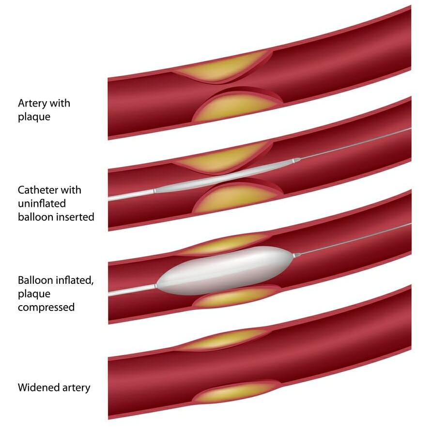
PVD is a condition that effects the arteries of the upper and lower limbs reducing blood circulation. It is produced by atherosclerosis which is the build up of fatty lipids (plaque) within the wall of arteries. This causes a narrowing (stenosis) or blockage in the artery and reduces blood flow to the area it supplies.
Causes and Symptoms of Peripheral Vascular Disease (PVD) Risk Factors:
OR Request an Appointment
Symptoms of PVD will depend on which artery is narrowed or blocked. This most commonly occurs in the arteries supplying the legs.
When a blockage of an artery to the leg occurs, it decreases blood supply to the muscles of the leg and this causes the muscles to become painful or to cramp on walking a certain distance. Pain in the muscles on exercise is called Claudication. Claudication is one of the first signs of PVD. Claudication can occur at any distance but often presents on walking about 400 metres. When the patient stops walking, the pain is relieved but recurs if they resume walking the same distance. If untreated this can progress to pain on walking even shorter distances.
Arterial disease can progress to pain in the leg at night, at which stage the patient may have to dangle their foot outside the bed to provide temporary pain relief. PVD if left untreated can progress to a stage where pain is present all the time, even at rest.
Severe PVD can lead to ulcers and gangrene. It is therefore important to see a doctor at the commencement of symptoms of PVD to prevent any progression of the disease to more serious outcomes.
Diagnosis of Peripheral Vascular Disease (PVD)
The diagnosis of PVD is made by a thorough patient history, physical examination and radiological investigation which determines the underlying causes and where the artery is blocked.
A non-invasive ultrasound evaluation of the arteries is the initial investigation undertaken to determine the position of the blockage and assists in the planning of treatment. Occasionally, a Computed Tomography (CT) angiogram scan is also used to help confirm the site of the blockage.
Treatment of Peripheral Vascular Disease (PVD)
The goal of treatment in PVD is to reduce symptoms by treating the underlying causes. This may involve stopping smoking, improving diet, having regular exercise, and taking medications to treat high blood pressure (hypertension), high cholesterol (hyperlipidemia) and diabetes if present.
The next stage is to improve the blood flow beyond the blocked artery.
In 90% of cases, the blood flow to the leg is improved with minimally invasive angioplasty and/or stenting procedures to open narrowed or blocked arteries. This technique involves the use of catheters, balloons and stents. It occurs through a small keyhole incision in the groin performed under local anaesthetic to access the femoral artery which allows access to unblock the diseased artery.
What is Angioplasty?
Angioplasty is a procedure using a catheter which incorporates a balloon to dilate the artery. Using image guidance the catheter is inserted through the artery to the area of the blockage. The balloon is inflated opening the artery and improving the blood flow. The balloon is then deflated and the catheter and balloon removed. Angioplasty may be used with or without stenting.
What is a stent?

Stents are small, expandable tubes which are inserted into the blocked arteries to mechanically support the structure of the arterial wall. The stent functions to hold the artery open and increase blood flow through it. Stents remain in the artery and act to prevent the artery narrowing again.
Angioplasty and stenting procedures are performed under local anaesthetic and sedation allowing most patients to go home the day of the procedure or the following day.
Occasionally because of significant arterial disease, open bypass surgery to replace or reroute the blood supply around the blocked part of the artery may still be required to improve the blood flow.


